click on one of the cell types below to be taken to its interactive 3D model
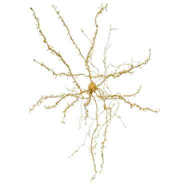
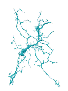

MSN
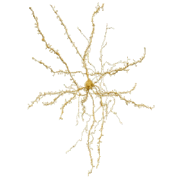
Medium spiny neurons (MSNs) are named after their numerous dendritic spines. They have analogues in the mammalian striatum which project to other basal ganglia nuclei, and are therefore also referred to as spiny projection neurons (SPNs). MSNs are part of the direct and indirect pathways. In our Area X dataset, MSNs are the smallest adult neuronal cell type, but also the most abundant: 94.4% of the adult neurons analysed were MSNs.
putative neurotransmitter: GABA
browse real data: here
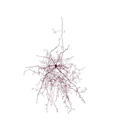
STN
Named after their potential mammalian analogues, the projection neurons of the subthalamic nucleus (STN) are the only glutamatergic projection neurons in the basal ganglia. STNs form part of the indirect and hyperdirect pathways. In our dataset, they account for 1.2% of adult neurons.
putative neurotransmitter: glutamate
browse real data: here
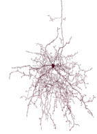
TAN
Tonically active neurons (TANs) are cholinergic interneurons (ChINs). They have mammalian analogues in the striatum. In our dataset, they have the largest somata, which are covered in spines and have large axonal arborizations. Due to their size, only a few TAN somata are included in the dataset, accounting for just 0.1% of adult neurons.
putative neurotransmitter: acetylcholine
GPe
Their putative mammalian analogues are the prototypical projection neurons in the globus pallidus externus (GPe). In the mammalian literature, these neurons are usually described as being part of the indirect pathway. However, in our dataset, their main projection targets lie outside this pathway (INT2 and INT3). Their dendrites contain very few spines, and GPe neurons account for just 0.3% of adult neurons.
putative neurotransmitter: GABA
browse real data: here
GPi
Their putative mammalian analogues are the projection neurons of the globus pallidus internus (GPi) and the substantia nigra pars reticulata (SNr). These neurons are tonically active with high firing rates and are basal ganglia output neurons. In our dataset, these cells have large somata, large axonal boutons, and thick, myelinated axon branches that exit the dataset. This indicates that they are indeed the neurons that project outside of Area X, accounting for 0.5% of adult neurons.
putative neurotransmitter: GABA
browse real data: here
LTS
These neurons are named after their low-threshold spikes (LTS) and are analogous to interneurons in the striatum. They have long, sparse axonal arborisations and make up 0.4 % of adult neurons in our dataset.
putative neurotransmitter: GABA
browse real data: here
INT1
This cell type is one of three newly described interneuron types in the dataset. With its dense axonal arborisations with regular boutons, it resembles the fast-spiking interneuron type found in the mammalian striatum in terms of morphology. However, closer analysis revealed that the three types differ in morphology and connectivity. In our dataset, INT1 accounts for 1.1% of adult neurons.
putative neurotransmitter: GABA
browse real data: here
INT2
This cell type is one of three newly described interneuron types in the dataset. Due to its dense axonal arborisations and regular boutons, its morphology is similar to that of the fast-spiking interneuron type found in the mammalian striatum. However, closer analysis revealed morphological and connectivity differences between the three types. Of the three newly described interneuron types, INT2 has the smallest soma and the greatest number of spines on its dendrites. INT2 accounts for 1.2% of adult neurons.
putative neurotransmitter: GABA
browse real data: here
INT3
This cell type is one of three newly described interneuron types in the dataset. Due to its dense axonal arborisations and regular boutons, its morphology is similar to that of the fast-spiking interneuron type found in the mammalian striatum. However, closer analysis revealed morphological and connectivity differences between the three types. Of these, INT3 has the largest soma and the longest axons. Additionally, half of the neurons form autapses, accounting for 0.8% of adult neurons.
putative neurotransmitter: GABA
Migratory neuron
Migratory neurons (MIGR) are immature neurons that migrate through the brain. Songbirds generate new neurons throughout life, which migrate through the brain before being recruited into song-relevant circuitry as mature neurons. In our dataset, MIGR have a simple morphology consisting of an elongated soma and an extending leading process, and are thought to travel in the direction of this leading process.
browse real data: here
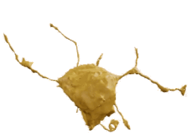
Astrocyte
Astrocytes are glial cells that control the environment within the brain and access to it by forming part of the blood–brain barrier. They have an elongated soma and several branches that form a complex tree structure with several thin processes at the end. At least one of their endfeet makes contact with a blood vessel, as can be seen in the example. One endfoot looks like a loop there. If you check the raw data, you will see that the loop encloses a blood vessel!
browse real data: here
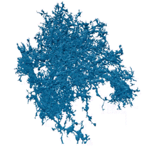
Microglia
Microglia are immune cells found in the brain. In our dataset, we observe them in their 'resting' state, during which they are actually surveilling the environment. At this stage, they have a small soma and several processes which split into thin branches. Once they are activated, their morphology changes and becomes more amoeboid, which is currently not classified in the dataset. In the current automatic classification, oligodendrocyte precursor cells (OPCs) are not classified as a distinct group, but are instead included in the microglia category.
browse real data: here
Oligodendrocyte (partial)
This glial cell type extends its plasma membrane to produce the myelin sheet that wraps around neurons, thereby enhancing axonal signal transmission. They can also provide metabolic support. Oligodendrocytes extend several thin processes in order to myelinate axons. Unfortunately, due to their thinness, these are often cut off in our dataset. As you can see in the example, one of the processes creates a loop. In the raw data, you can see that this is where the process connects to the myelin sheet wrapped around a GPi axon.
browse real data: here
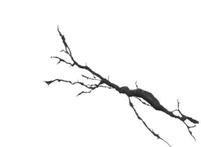
DA
This cell type comprises putative dopaminergic axons which project from the ventral tegmental area (VTA) to Area X. While these axons have a low synapse density, they have a high density of vesicle clouds along the axon. In the current model of song learning, dopamine signals reward prediction or performance error.
putative neurotransmitter: dopamine
browse real data: here
HVC
This cell type comprises putative axons from the cortical (pallial) region HVC (used as a proper name). In the current model of song learning, HVC axons provide a timing signal.
putative neurotransmitter: glutamate
browse real data: here
LMAN
This cell type comprises putative axons from the lateral magnocellular nucleus of the anterior nidopallium (LMAN) in the cortex (pallium). In the current model of song learning, LMAN axons provide a variability signal that is used to explore different song variations during the learning process.
putative neurotransmitter: glutamate
Check out our interactive 3D models at the below links